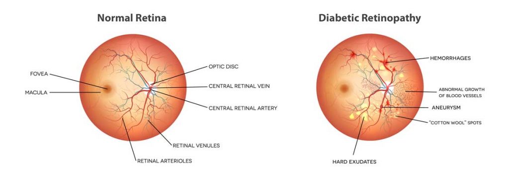
The eye disease diabetic retinopathy is the fastest growing cause of blindness in the world. There are over 100 million people in the world with diabetes and ideally, they would be screened each year for this degenerative eye condition. It is fully treatable if caught early, however if it’s not detected you could suffer partial or full vision loss. When an ophthalmologist studies an eye image, they grade it on a five-point scale, one through five. They look for scattered hemorrhages and micro-aneurysms to grade the image. This is quite subjective in that a grade of two (2) means come back in a year and a grade of three (3) means come to the clinic right away. We all know second opinions in medicine are often recommended. Studies have shown however, if you ask two ophthalmologists to grade the same retinal image, they will agree only 60% of the time. Image analysis in radiology and other medical areas are quite subjective. Fortunately, machines can be trained to read and interpret image data.
Deep Learning can enable a predictive analytical approach to healthcare.
A data set of 128,175 retinal images was used to train a deep convolutional neural network using a retrospective development algorithm. According to the Journal of the American Medical Association (JAMA) article, “the algorithm had 90.3% and 87.0% sensitivity and 98.1% and 98.5% specificity for detecting referable diabetic retinopathy, defined as moderate or worse diabetic retinopathy or referable macular edema by the majority decision of a panel of at least 7 US board-certified ophthalmologists.” In medical diagnosis, test sensitivity is the ability of a test to correctly identify those with the disease (true positive rate), whereas test specificity is the ability of the test to correctly identify those without the disease (true negative rate). A 90% prediction that something is wrong with your eye is quite good!

Now the researchers at Google thought to take this further by getting images labeled by specialists who have more training in retinal eye disease. Rather than using independent assessments, or singular data points, they got three retinal specialists in a room to come up with an adjudicated number for each image. Google researchers then used that data to train the deep learning model further and were able to achieve prediction outcomes on par with what we’d expect from highly trained specialists. This all makes the point of why the data preparation step is so important in machine learning development.
Solving one problem with Deep Learning often enables discovery of other use cases.
By taking the model and adding more training parameters and convolutional layers to the neural network, you can now predict many other things with a retinal image. Predictions can be made about a person’s age, their gender, their hemoglobin level and their systolic and diastolic blood pressure. When you combine all of this together you can get a prediction of someone’s cardiovascular risk – at the same level of accuracy that normally requires a more invasive blood test. The first approved instrument to provide a screening decision without clinician input was granted by the FDA to the cloud-based software system IDx-DR (IDx Technologies) in 2018. This medical device can detect referable diabetic retinopathy (DR) from color fundus photographs.
In India, there is a shortage of more than 100,000 eye doctors to do the necessary screening of diabetic retinopathy and so 45% of patients endure vision loss before they’re diagnosed. This is tragic. Using AI, and more specifically deep learning, we can enable greater reach into population groups with low cost imaging equipment where doctors are in short supply. Having a longitudinal history of the eye that can be analyzed remotely, can not only prevent blindness, but also other detect other health conditions as well.
References: Jama Network article 2588763, Research Google pub45732, Nature article s41746-018-0040-6
